Radius
Except in immature individuals, the radius and ulna are commonly fused near their distal ends. This “articulation” or lack of it is ignored for the purpose of analysis and each element is analyzed separately. Fused and isolated distal ends of ulna may or may not be present on individual radii analyzed.
Elements where the proximal and/or distal epiphyses are un-fused and not with the specimen are coded as though the epiphyses are missing.
Elements where the proximal or distal epiphyses are un-fused but are present during coding, the un-fused epiphyses are grouped together for coding and cataloguing and are not counted as separated pieces.
For an un-fused epiphysis unassociated with the rest of the element it is assigned an appropriate BU number.
For the Fincastle Project, Bone Unit 24 was redefined, and Bone Unit 34 was added.
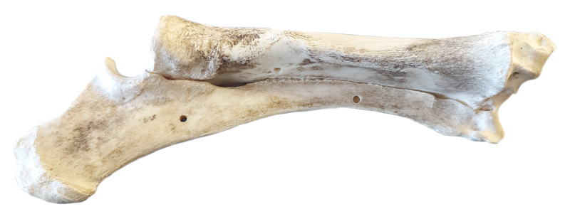
BU 1
SC A
Complete element.
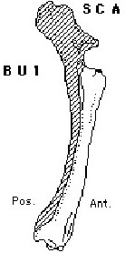
BU 14
SC B
Unit consists of complete proximal end and ¾ or more or adjoining shaft. Distal end completely absent.
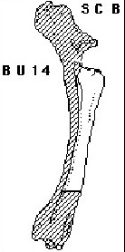
BU 10
SC C
Complete proximal end of radius with ¼ - ½ of adjoining shaft represented.
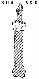
BU 3
SC D
Complete proximal end of radius with 0 – ¼ of shaft length represented.
BU 29
SC E
Unit consists of the lateral half of the element only, split sagittally. (WU)

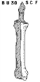
BU 30
SC F
Unit consists of medial half of the element, split sagittally. (WU)
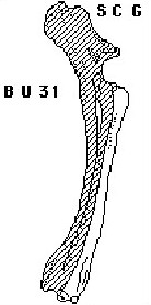
BU 31
SC G
Unit consists of anterior section of the entire element (from proximal to distal end), split medially. (TVH)
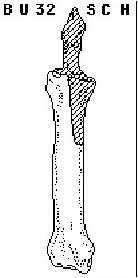
BU 32
SC H
Unit is missing the proximal medial articular surface and up to ½ the adjoining medial shaft. Opposite of BU 9. (WU)
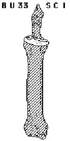
BU 33
SC I
Unit consists of unfused proximal epiphysis. Coded as ‘weathered’. (WU)
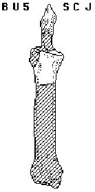
BU 5
SC J
Proximal end of radius missing only a small portion of the smaller lateral articular facet. 0 – ¼ of shaft length represented.
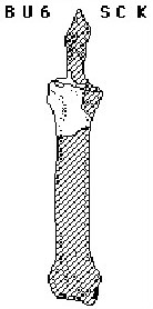
BU 6
SC K
Proximal end of radius missing only a small portion of the larger lateral articular facet. 0 – ¼ of shaft length represented.
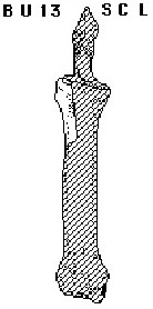
BU 13
SC L
Fragment of proximal end consisting of much or all of lateral articular surface. 0 – ¼ of total shaft length represented.
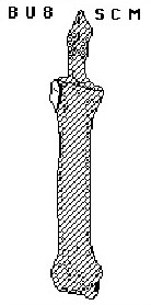
BU 8
SC M
Large to small portion of shaft including small portion of lateral articular facet at proximal end. Similar to BU 13.
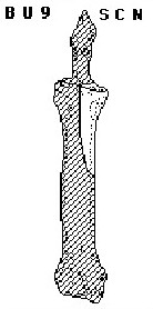
BU 9
SC N
Large portion of proximal end consisting of all or most of large medial articular surface. Between ¼ - ½ of adjoining shaft represented.
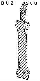
BU 21
SC O
All or most of medial articular surface at proximal end with from 0 – ¼ of adjoining shaft represented. Similar to BU 9 but with less shaft. Similar to BU 7. (TH)
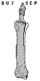
BU 7
SC P
Large to small section of shaft including a small portion of the larger medial articular facet at proximal end. Medial portion of rough articular scar from contact with ulna may also be present. Similar to BU 21.
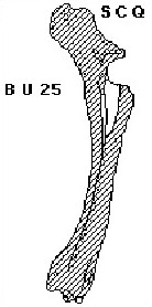
BU 25
SC Q
Posterior half of the proximal end, with 0 – ¼ of the adjoining shaft represented (posterior half of BU 3). (WU)
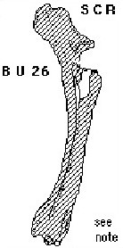
BU 26
SC R
Posterior half of the proximal medial end, missing the lateral articular facet and the anterior half of the entire end. 0 – ¼ if the adjoining shaft represented (posterior half of BU 5). (WU)
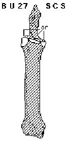
BU 27
SC S
Fragments from the proximal end of the radius. Similar to BU 7, BU 8, BU 12, BU 13 and BU 15 but indeterminate as to which. (WU) (TH)
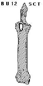
BU 12
SC T
Small fragment of proximal end characterized by presence of coronoid process. Very little of shaft or articular condyles present. Similar to BU 15.
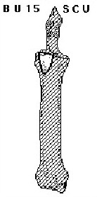
BU 15
SC U
Small fragment of proximal end characterized by presence of coronoid process and small to moderate portions of the adjoining medial and lateral condyles. Similar to BU 12.
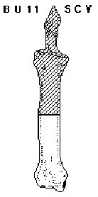
BU 11
SC V
Complete distal end of radius and between ¼ - ½ of adjoining shaft represented.
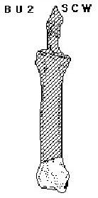
BU 2
SC W
Complete distal end of radius with 0 – ¼ of adjoining shaft length. Also commonly includes distal end of ulna fused to unit.
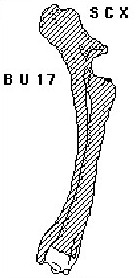
BU 17
SC X
Unfused, distal end of radius detached and not associated with rest of element. Condition coded as ‘weathered’.
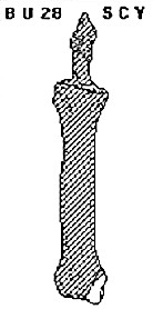
BU 28
SC Y
Broken medial half of an unfused distal epiphysis. Coded as ‘weathered’. Similar to BU 22. (WU)
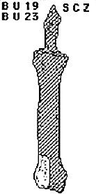
BU 19
SC Z
Lateral half of distal end with from 0 – ¼ of adjoining shaft represented. May have distal end of ulna attached. BU 23 was also assigned to this element configuration. Either BU number can be used in coding. (TH)

BU 23
SC Z
Lateral half of distal end with from 0 – ¼ of adjoining shaft represented. May have distal end of ulna attached. BU 23 was also assigned to this element configuration. Either BU number can be used in coding. (TH)
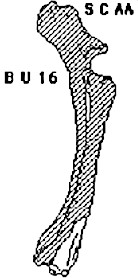
BU 16
SC AA
Anterior portion of distal end with articular surface and 0 – 1/3 of adjoining shaft represented. (TH)
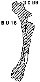
BU 18
SC BB
Medial to distal fragment of shaft from along anterior surface. Identifiable on basis of characteristic shape and form. (TH)
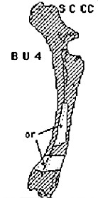
BU 4
SC CC
Small to large fragment of shaft characterized by small segment of ulna fused to it or simply portion of rough scarred surface adjoining unfused section of ulna. Frequency indicates number of pieces only. Nutrient foramen may or may not be present.
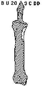
BU 20
SC DD
Proximal, medial fragment of shaft characterized by characteristic cross-section or muscular scarring. Lacks any of proximal end. (TH)
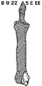
BU 22
SC EE
Distal medial articular surface, may include portion of shaft. (EMA)
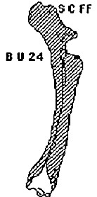
BU 24
SC FF
Posterior portion of distal end with articular surface and 0 – 1/3 of adjoining shaft represented. May have portions of ulna fused to it.
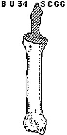
BU 34
SC GG
Complete juvenile missing proximal epiphysis. Opposite BU 33. (SB)