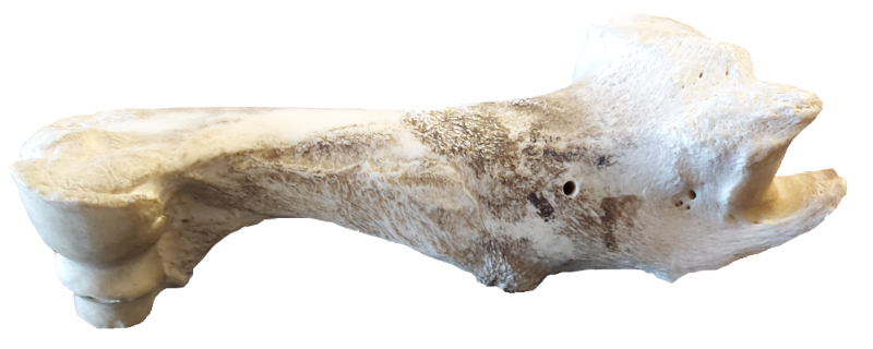
Humerus
Elements where the proximal and/or distal epiphyses are un-fused and not with the specimen are coded as though the epiphyses are missing.
Elements where the proximal or distal epiphyses are un-fused but are present during coding, the un-fused epiphyses are grouped together for coding and cataloguing and are not counted as separated pieces.
For an un-fused epiphysis unassociated with the rest of the element it is assigned an appropriate BU number.
For the Fincastle Project, Bone Units 39 and 40 were added.
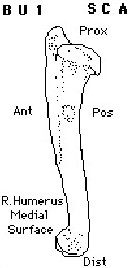
BU 1
SC A
Complete element.
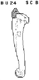
BU 24
SC B
Consists of nearly complete element lacking only the lateral tuberosity. (AL)
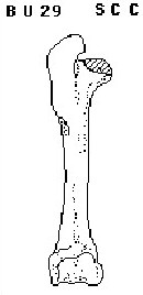
BU 29
SC C
Complete humerus except for absence of medial tuberosity and adjoining intertuberal groove.
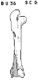
BU 36
SC D
Element is complete, with exception of a missing lateral distal condyle. This unit is the opposite of BU 4. (WU)
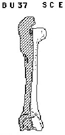
BU 37
SC E
Unit consists of only the medial half of the element. Missing portions include the proximal lateral tuberosity and the distal lateral condyle, as well as all lateral portions of the shaft. (WU)
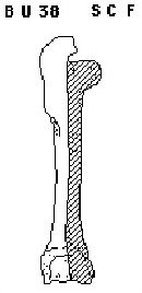
BU 38
SC F
Unit consists of only the lateral half of the element. Missing portions include the proximal medial tuberosity and the distal medial condyle, as well as all medial portions of the shaft. (WU)
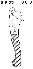
BU 33
SC G
Complete proximal end of humerus with ¼ - ½ of adjoining shaft. (KK)
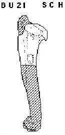
BU 21
SC H
Complete proximal end of element with ¼ - ½ of shaft represented. Lateral tuberosity absent. Tuberosity absent due to either weathering or lack of fusion. (AL)
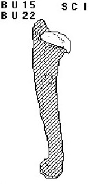
BU 15
SC I
Complete humeral head, unfused and detached from rest of element. Margins of unit often gnawed or battered. Differentiated from BU 5 by absence of any adjoining articular surfaces of humerus and fact that it has not been detached by butchering. Observation not made on Cactus Flower material analyzed in 1974. BU 22 was also assigned to this element configuration. Either BU number can be used in coding. (JB)

BU 22
SC I
Complete humeral head, unfused and detached from rest of element. Margins of unit often gnawed or battered. Differentiated from BU 5 by absence of any adjoining articular surfaces of humerus and fact that it has not been detached by butchering. Observation not made on Cactus Flower material analyzed in 1974. BU 22 was also assigned to this element configuration. Either BU number can be used in coding. (JB)
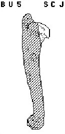
BU 5
SC J
Portion of head of humerus and often immediately adjoining portion of neck. Similar to BU 15.
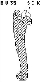
BU 35
SC K
Fragments of humeral head. Similar to BU 28. (WU)
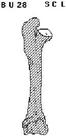
BU 28
SC L
Portion of medial tuberosity and intertuberal groove. (TH)
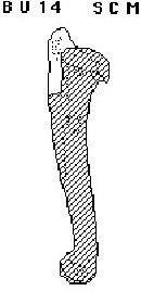
BU 14
SC M
Small to large section of shaft including all or a major portion of the lateral tuberosity.
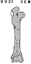
BU 27
SC N
Proximal shaft fragment along lateral surface characterized by rough prominence for attachment of infraspinatus tendon. Immediately below or distal to lateral tuberosity. (JB)
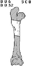
BU 6
SC O
Small to large fragment of shaft, identifiable by presence of the deltoid tuberosity. BU 32was also assigned to this element configuration. Either BU number can be used in coding. (JB)

BU 32
SC O
Small to large fragment of shaft, identifiable by presence of the deltoid tuberosity. BU 32was also assigned to this element configuration. Either BU number can be used in coding. (JB)
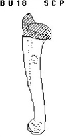
BU 18
SC P
Element with head and lateral tuberosity removed; extreme posterior margin of head may or may not be present. Three quarters or more of shaft and entire distal end present.
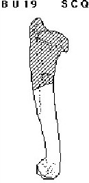
BU 19
SC Q
Complete distal end of humerus with ½ - ¾ of adjoining shaft length represented. Similar to BU 16 an BU 18.
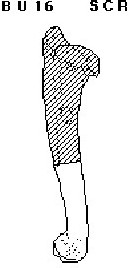
BU 16
SC R
Complete distal end of humerus with ¼ - ½ of adjoining shaft length represented. Similar to BU 19.
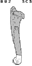
BU 2
SC S
Complete distal end of humerus and 0 – ¼ of adjoining shaft length.
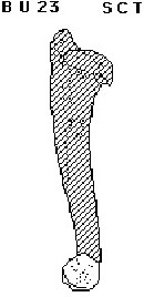
BU 23
SC T
Unfused, complete or more of half of distal epiphyses detached and not associated with rest of element. Condition coded as ‘weathered’.
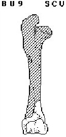
BU 9
SC U
Complete distal end of humerus with from 0 – ¼ of shaft represented. A small to large fragment of shaft from along the lateral edge immediately above the lateral condyles (BU 7) is missing. Similar to BU 2 and BU 10.
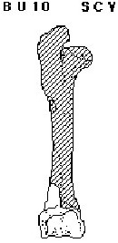
BU 10
SC V
Complete distal end of humerus with from 0 – ¼ of shaft represented. A small to large fragment of shaft from along the medial edge immediately above the medial condyles (BU 8) is missing. Similar to BU 2 and BU 9.
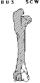
BU 3
SC W
Medial half of distal end of humerus. Consists of all or most of medial epicondyle and 0 – ¼ of adjoining shaft length.
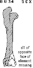
BU 34
SC X
Anterior portion of the medial half of the distal end of the element, with 0 – ¼ of the adjoining shaft (the anterior half of BU 3). (WU)
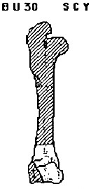
BU 30
SC Y
Distal end of humerus with 0 – ¼ of adjoining shaft represented. Lateral epicondyle missing. (KK)
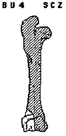
BU 4
SC Z
Lateral half of distal end of humerus. Consists of all or most of lateral epicondyle and 0 – ¼ of adjoining shaft length.
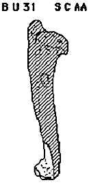
BU 31
SC AA
Medial and lateral condyles with medial and lateral epicondyles removed. May include 0 – ¼ of adjoining shaft. (KK)
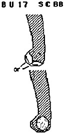
BU 17
SC BB
Shattered pieces of distal end of humerus identifiable by distinctive surfaces of either the lateral or medial epicondyle. Pieces usually too incomplete to identify as being either the medial or lateral epicondyle; or left or right; and as to minimum number of elements represented. Frequency indicates number of pieces only.
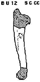
BU 12
SC CC
Large section of shaft from which proximal and distal ends have been completely removed. Usually this large piece is a section of ‘tube’. Both deltoid tuberosity and teres tubercle present.
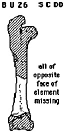
BU 26
SC DD
Distal anterior portion of shaft above and containing part of coronoid fossa. Does not include any of olecranon fossa. May contain portions of one or both lateral margins. Similar to BU 7 and BU 8 combined. (TH)
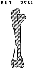
BU 7
SC EE
Small to large fragment of shaft from along the lateral edge immediately above the lateral condyles. Unit identifiable because it includes part of the coronoid fossa and/or the olecranon fossa. Similar to BU 8 and BU 11.
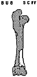
BU 8
SC FF
Small to large fragment of shaft from along the medial edge immediately above the medial condyles. Unit identifiable because it includes part of the coronoid fossa and/or the olecranon fossa. Similar to BU 7 and BU 11.

BU 11
SC GG
Either BU 7 or BU 8. Indeterminate as to which.
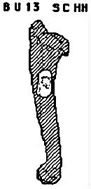
BU 13
SC HH
Small to large section of shaft characterized by presence of teres tubercle.
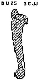
BU 25
SC JJ
Posterior section of shaft above olecranon fossa. Up to 1/3 of shaft length represented and exhibits beginning of olecranon fossa. Nutrient foramen not represented. Similar to BU 20. (TH)
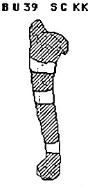
BU 39
SC KK
Shaft fragments identifiable by shape and curvature. (SB)
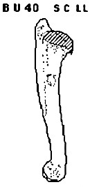
BU 40
SC LL
Complete juvenile element, missing the proximal epiphysis only. Opposite from BU 15/22. (SB)