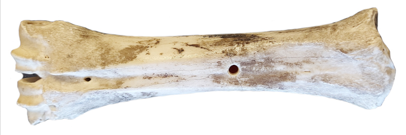
Metatarsal
Elements where the proximal and/or distal epiphyses are un-fused and not with the specimen are coded as though the epiphyses are missing.
Elements where the proximal or distal epiphyses are un-fused but are present during coding, the un-fused epiphyses are grouped together for coding and cataloguing and are not counted as separated pieces.
For an un-fused epiphysis unassociated with the rest of the element it is assigned an appropriate BU number.
For the Fincastle Project, Bone Units 16, 17 and 18 were added.
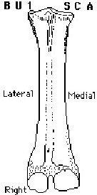
BU 1
SC A
Complete element.
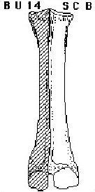
BU 14
SC B
Unit consists of one half of element (either medial or lateral side) split along the sagittal plane. (WU)
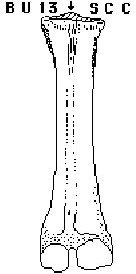
BU 13
SC C
Element missing all or most of posterior edge of proximal end. Incompletely illustrated. (GL)
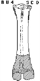
BU 4
SC D
Complete proximal end with 1/2 – 3/4 of shaft represented.
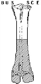
BU 3
SC E
Complete proximal end with 1/4 – 1/2 of shaft represented.
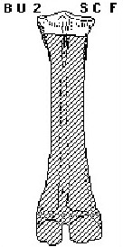
BU 2
SC F
Complete proximal end with 0 – 1/4 of shaft represented.
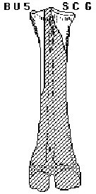
BU 5
SC G
Shattered proximal end pieces. Proximal end broken into two or more pieces of various sizes and configurations. From 0 – 3/4 shaft length may be represented; most commonly 1/4 – 1/3. Frequency indicates minimum number of elements represented by pieces. For Oldman project, frequency indicates number of pieces only.

BU 6
SC H
Not a butchering unit. Frequency indicates number of pieces constituting BU 5. Not coded for Oldman unit.
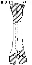
BU 11
SC I
Shattered shaft fragment characterized by vascular groove and commonly vascular foramen. Greater than 1/2 total shaft length represented. None of distal epiphyses present. Frequency indicates minimum number of elements.
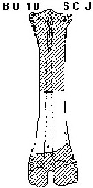
BU 10
SC J
Shattered shaft fragment characterized by vascular groove and vascular foramen. No more than 1/2 total shaft length represented. None of the distal epiphyses present. Frequency indicates minimum number of elements.
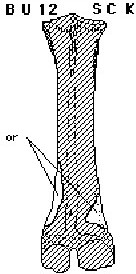
BU 12
SC K
Shattered shaft fragment. Characterized by shaft shape. Lacks vascular groove. (TH)
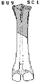
BU 9
SC L
Complete distal end with 1/2 – 3/4 of shaft length represented. Similar to BU 7. (SB)
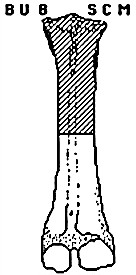
BU 8
SC M
Same as BU 7 but with approximately ¼ - ½ of distal shaft length represented.
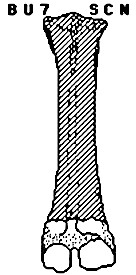
BU 7
SC N
Complete distal end of metatarsal with 0 – ¼ of adjoining distal shaft represented. Note: many distal ends from either metacarpals or metatarsals analyzed as separate element under metapodial fragments.
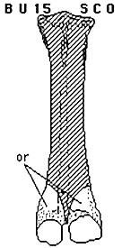
BU 15
SC O
Unit consists of one of either distal condyles and from 0 – ¼ of the adjoining shaft. The unit is similar to metapodial BU 4, but the element is identifiable to the specific metapodial. (WU)
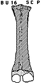
BU 16
SC P
Distal epiphysis only (juvenile). (SB)
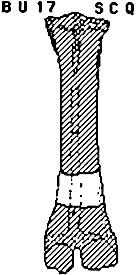
BU 17
SC Q
Similar to BU 12, shaft fragment but has vascular groove. (SB)
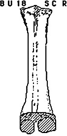
BU 18
SC R
Proximal end with the entire shaft; missing the distal epiphysis (juvenile). (SB)