Femur
Elements where the proximal and/or distal epiphyses are un-fused and not with the specimen are coded as though the epiphyses are missing.
Elements where the proximal or distal epiphyses are un-fused but are present during coding, the un-fused epiphyses are grouped together for coding and cataloguing and are not counted as separated pieces.
For an un-fused epiphysis unassociated with the rest of the element it is assigned an appropriate BU number.
For the Fincastle Project, Bone Units 34 to 40 were added.
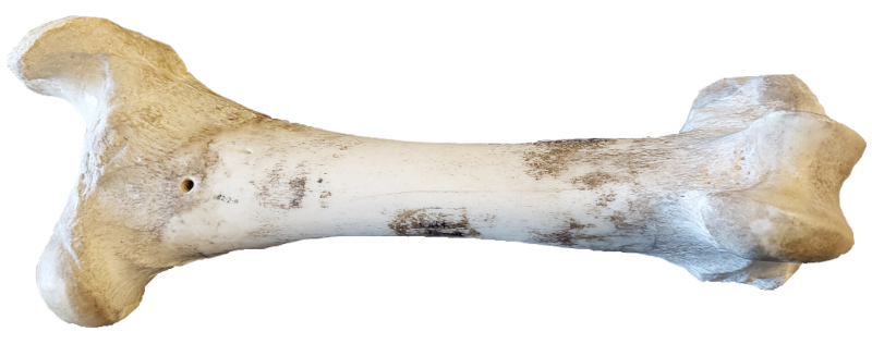
BU 1
SC A
Complete element.
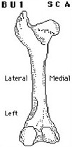
BU 30
SC B
Unit is essentially a complete femur, with the exception of a missing femoral head and neck. Missing due to weathering or butchering – not lack of fusion. Opposite of BU 2. (TVH)
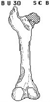
BU 16
SC C
Essentially complete femur missing greater trochanter as a result of butchering or weathering and not due to lack of epiphyseal fusion. Similar to BU 19.
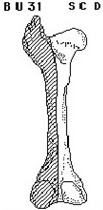
BU 31
SC D
Unit consists of the medial portion of the element only, split along the sagittal plane. Present distal medial epicondyle, medial shaft, and proximally, the trochanter minor. The lateral half of the shaft, distal lateral epicondyle and proximal articular surfaces are not present. (THV)
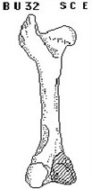
BU 32
SC E
Unit is essentially complete, with the exception of a missing medial condyle. Opposite of BU 6. (WU)
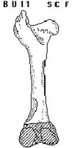
BU 11
SC F
Unit consists of complete proximal end and virtually complete shaft; supracondolid fossa present on shaft. Distal end completely absent due to butchering or weathering.
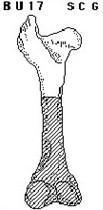
BU 17
SC G
Complete proximal end of element with proximal ¼ - ½ of shaft represented.
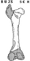
BU 23
SC H
Proximal end of element missing greater trochanter. From all to most of adjoining shaft represented. All or most of supracondyloid fossa represented. (TH)
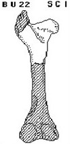
BU 22
SC I
Proximal end of element missing greater trochanter with proximal ¼ - ½ of shaft represented. (AL)
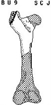
BU 9
SC J
All or most of trochanter major, trochanteric fossa and trochanter minor (rest of trochanter major shows light to extensive damage either as a result of crushing or carnivore gnawing- indeterminate as to which).
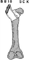
BU 10
SC K
Same as BU 9 but trochanter minor not included.
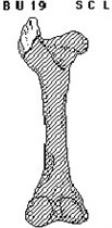
BU 19
SC L
Unfused greater trochanter detached and not associated with rest of element. Code condition as ‘weathered’. Similar to BU 6.
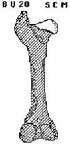
BU 20
SC M
Unfused femoral head detached and not associated with rest of element. Code condition as ‘weathered’. Similar to BU 2.
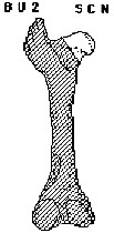
BU 2
SC N
All or major portion of femoral head. Small to moderate portion of neck commonly present. Similar to BU 24.
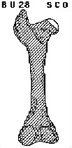
BU 28
SC O
Fragment of femoral head, characterized by the presence of the fovea capitis. Similar to BU 2.
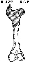
BU 29
SC P
Complete distal femur with ½ - ¾ of adjoining shaft. (WU)
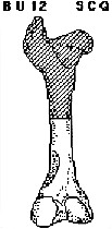
BU 12
SC Q
Unit consists of complete distal end and ¼ - ½ of the shaft.
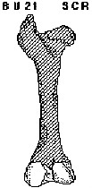
BU 21
SC R
Unfused distal epiphyses detached and not associated with rest of element. Code condition as ‘weathered’.
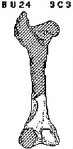
BU 24
SC S
Distal end with up to half of distal end of shaft represented. All or major portion of supracondyloid fossa represented. Lateral epicondyle removed. (KK)
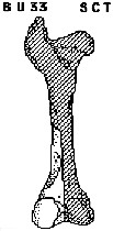
BU 33
SC T
Unit consists of the lateral condyle and epicondyle, and a portion of the adjoining shaft, including the supracondyloid fossa. (WU)
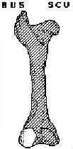
BU 5
SC U
All or major portion of lateral epicondyle detached from rest of element.
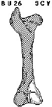
BU 26
SC V
Medial epicondyle with 0 – ½ of adjoining shaft represented. Similar to BU 6. (KK)
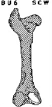
BU 6
SC W
All or most of medial epicondyle detached from rest of element. Similar to BU 26.
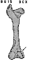
BU 15
SC X
Large to small portion of medial epicondyle at distal end of shaft identifiable by distinctive shape configuration and muscular scarring.
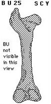
BU 25
SC Y
Trochlea of distal anterior surface (not visible in drawing). (GL)
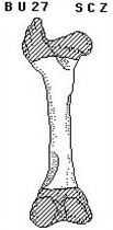
BU 27
SC Z
Complete femoral shaft with head, greater trochanter and distal end missing. (KK)
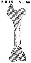
BU 13
SC AA
Large fragment of shaft characterized by presence of all or a major portion of both the trochanter minor and the supracondaloid fossa. The fragment is not enclosed or ‘tube-like’.
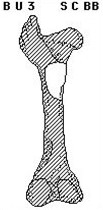
BU 3
SC BB
All or major portion of trochanter minor with a small to moderate amount of adjoining shaft present. The fragment is not enclosed or ‘tube-like’. Similar to BU 7.
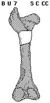
BU 7
SC CC
Medium to large ‘tube-like’ segment of shaft characterized by presence of trochanter minor. Similar to BU 3.
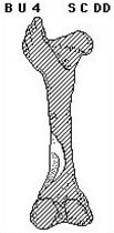
BU 4
SC DD
All or major portion of supracondyloid fossa with small to large amount of adjoining shaft length represented. The fragment is not enclosed or ‘tube-like’. Frequency indicates MNE, except for the Oldman Dam project, where frequency indicates the number of pieces only.
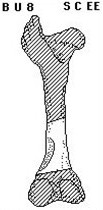
BU 8
SC EE
Moderate to large sized ‘tube-like’ segment of shaft characterized by presence of supracondyloid fossa.
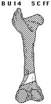
BU 14
SC FF
Small portion, no more than ½ of supracondyloid fossa, with small to moderate amount of adjoining shaft represented. Similar to BU 4. Frequency indicates MNE, except for the Oldman Dam project, where frequency indicates the number of pieces only.
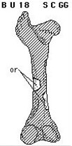
BU 18
SC GG
Fragments of femoral shaft, identifiable on basis of cross sectional shape. No portion of supracondyloid fossa or trochanter minor present.
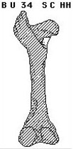
BU 34
SC HH
Complete or portion of the lesser trochanter ridge, detached from the condyles. (SB)
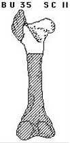
BU 35
SC II
Proximal end, but missing the greater trochanter with only 0 – 1/4 of shaft represented. Similar to BU 22. (SB)
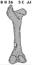
BU 36
SC JJ
Fragment of the shaft with a small portion of the supracondyloid fossa present. (SB)
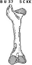
BU 37
SC KK
Juvenile element missing both the proximal and distal epiphyses. (SB)
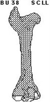
BU 38
SC LL
Fragment of the distal end identified by shape. (SB)
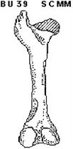
BU 39
SC MM
Juvenile missing the proximal epiphysis. Opposite BU20. (SB)
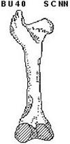
BU 40
SC NN
Juvenile missing distal epiphysis. Opposite BU 21. (SB)